Top research proposals to be offered equipment grants
The Mustafa(Pbuh) Science and Technology Foundation (MSTF) held a webinar on Application of Extracellular Recording Techniques in Neuroscience Research.
MSTF Media reports:
The second webinar of Innovation Lab webinars on neuroscience and electrophysiology, organized jointly by the Mustafa
Science and Technology Foundation and ScienceBeam Institute, was held virtually.
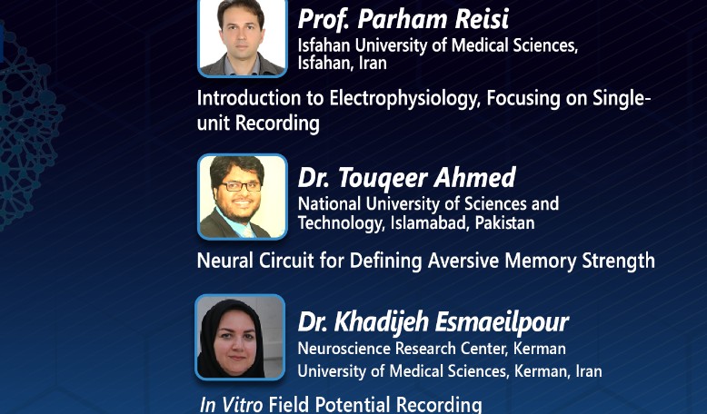
Three keynote speakers from different scientific centers gave lectures during the webinar.
Parham Reisi, Professor of Physiology at Isfahan University of Medical Sciences, first gave an introduction to electrophysiology, and then concentrated on single-unit recording.
Pointing to some basic aspects of electrophysiology, he defined electrophysiology as “the study of the electrical properties of biological cells and tissues.”
Electrical activity of cells, he pointed out, generates two main types of signals that reflect “either gradual changes in the membrane potential or all-or-none events.”
Electrophysiology involves measurements of voltage change or electrical current flow on a wide variety of scales from single ion channel proteins to whole tissues like the heart, he added.
Any electrical activity in the cell, he said, reflects a function in that cell.
Neurons are specialized for integration and propagation of electrical events, Reisi continued, adding that “It is through such electrical activity that neurons communicate with each other.” Therefore, an understanding of basic electrophysiology helps to recognize the function and dysfunctions of neurons.
Single-unit recording can be done both in vivo (on living animal) and in vitro (on brain slice), he said, adding that in vivo single-unit recording can be carried out either on anesthetized animal or freely moving animal.
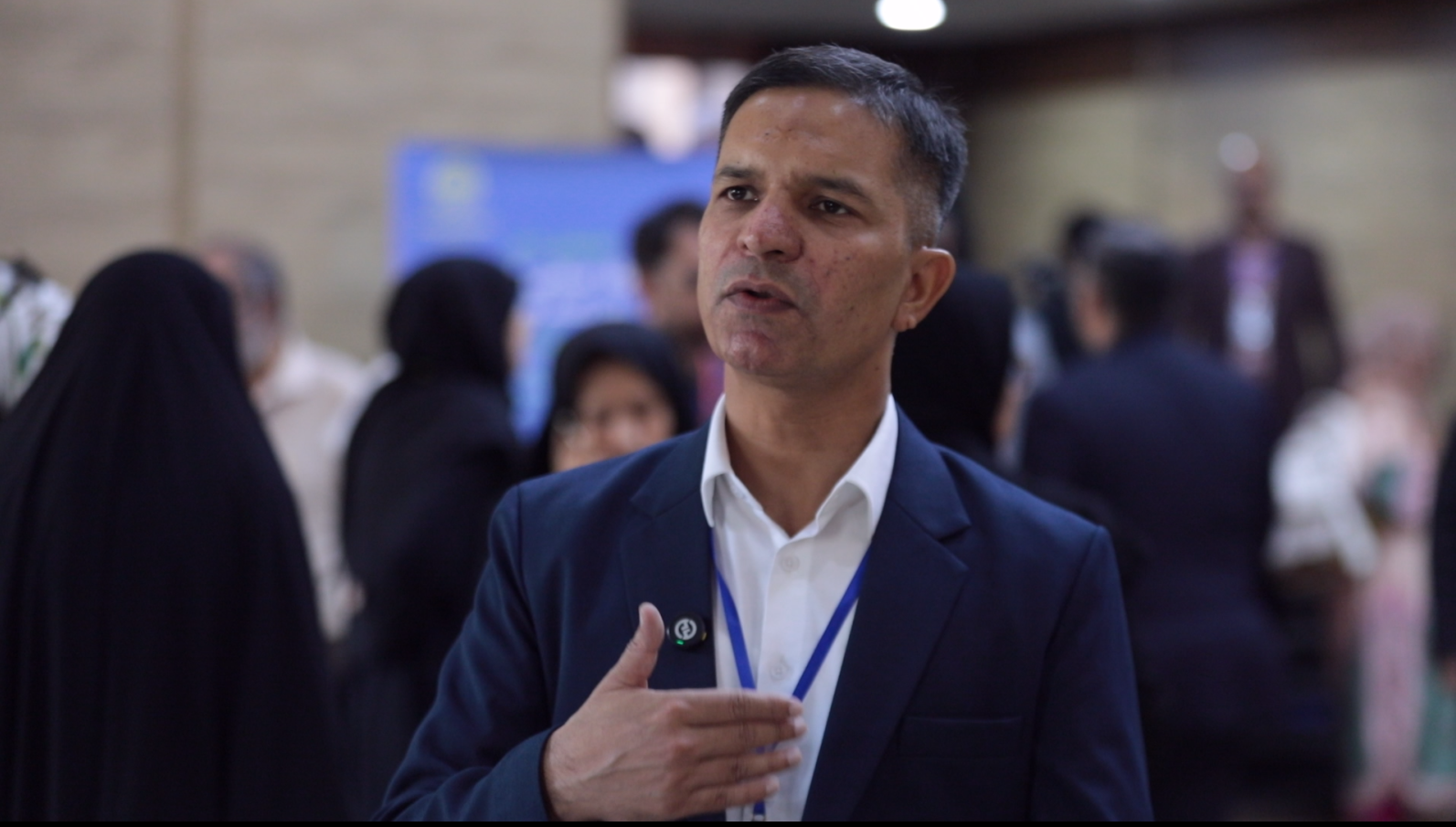
The next lecturer, Touqeer Ahmed, Associate Professor at National University of Sciences and Technology, Pakistan, delivered a speech on “Neural Circuit for Defining Aversive Memory Strength.”
“It is very instinctive to have fear memories. Whenever we see an aversive stimulus that stimulates aversive memory circuit and we get scared, a freezing response is produced that we actually hold there for some time,” he said.
In fear memories, a part of the brain called amygdala plays a crucial role, Ahmed said, adding that there are so many different areas in amygdala. He highlighted that his lecture mainly revolves around Central Amygdala (CeA) and Lateral Amygdala (LA).
“Aversive or fearful experiences powerfully regulate memory formation,” he noted, adding that “memory strength is proportional to the intensity of these experiences.”
Ahmed also talked about the strategy to study fear learning and strength, amygdala connections and fear learning circuit, and Lateral Amygdala (LA) neural response to shock.
He finally shared the conclusions of his study: Learning strength is regulated by feedback circuit of CeA-vIPAG and activation of LA neurons; CS processing is reduced in vIPAG neurons with optical inhibition of the CeA-vIPAG pathway; Fear-inducing auditory cues activate a vIPAG-RVM pain modulatory circuit that controls memory strength.
“It is possible that dysregulation of this feedback circuits in humans could lead to heightened and persistent fear memories leading to anxiety disorders,” Ahmed noted.
Khadijeh Esmaeilpour from Neuroscience Research Center at Kerman University of Medical Sciences, delivered a lecture on “In-Vitro Field Potential Recording Laboratory” during the webinar.
She briefly talked about electrophysiology, and then pointed to two types of electrophysiological recordings: Intracellular Recordings and Extracellular Recordings.
In intracellular recording, she said, electrode is placed into a single cell, while in extracellular recording the electrode is placed in extracellular space.
Pointing out that extracellular recording is divided into field potential recording, single-unit recording, and multi-unit recording, Esmaeilpour explained how each works.
Field potential recording is divided into two branches: in vitro, done on freshly sliced tissue—on which her presentation was chiefly focused—and in vivo, done on anesthetized animal.
Hinting at the advantages of in vitro method, she said in this approach “the extracellular space is accessible to the medium.” Moreover, there is the possibility of “direct visualization” and “rapid preparation.”
“The mechanical stability of preparation and simple control is another advantage of it,” she added.
She then explained how the slices are prepared in this method by showing a video clip to the audience, going through the stages one by one.
At the end of her lecture, Esmaeilpour pointed to the two papers she has published with her team on this topic: “Intranasal oxytocin administration facilitates the induction of long-term potentiation and promotes cognitive performance of maternally separated rats” and “Maternal separation impairs long-term potentiation in CA3-CA1 synapses in adolescent female rats.”
The Innovation Lab aims to develop neuroscience and electrophysiology research in the Islamic world by holding a series of webinars, a spring school, and offering grants. The first webinar, held in March with the collaboration of Pakistan’s Dow University of Health Sciences, revolved around “EEG and Neurofeedback Research and Clinical Applications.”
Supporting neuroscience and electrophysiology research in three areas including animal electrophysiology extracellular recording system (eLab/ ePulse); 4-128 channels EEG/QEEG/ERP acquisition system (eWave+); and neurofeedback and biofeedback system (eWave), 40,000 EUR equipment grants are offered to the best research proposals. The deadline to submit proposals is May 20, 2021. For more information, the candidates can send an email to
step@mstfdn.org and visit
www.sciencebeam.com.